Ventriculomegaly
Ventriculomegaly is a condition in which the fluid-filled spaces within the brain (ventricles) appear larger than normal on a fetal ultrasound, suggesting an increase in fluid in the fetal brain. Ventriculomegaly can often be confused with hydrocephaly, which is diagnosed after birth and is related to signs of increased pressure in the brain due to the increased fluid. Ventriculomegaly occurs in approximately 2 out of every 1,000 live births in the United States and can be identified on a fetal ultrasound as early as the second trimester.
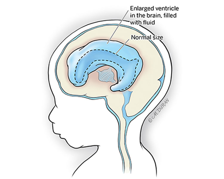
About Ventriculomegaly
Your child’s brain and spinal cord are covered in clear protective liquid known as cerebrospinal fluid. The spaces within the brain (ventricles) are also filled with cerebrospinal fluid. Produced in the brain’s ventricles, cerebrospinal fluid circulates around the brain and spinal cord and is absorbed through blood vessels and eventually filtered out of the body. In the brain of a healthy fetus, the ventricles are about 10 millimeters wide; however, cerebrospinal fluid can become trapped in the ventricles, causing the ventricles to expand and put extra pressure on the brain. When there is evidence of increased fluid in the fetal brain, the diagnosis of ventriculomegaly is made.
Ventriculomegaly can be associated with long-term neurologic developmental problems. In mild cases, more than 90% of children with ventriculomegaly will have a normal outcome. The incidence of developmental problems increases with the severity of the ventriculomegaly. Ventriculomegaly due to chromosomal problems and infections often has a poor prognosis.
Symptoms of Ventriculomegaly
Infants with mild self-limited (resolves on its own) ventriculomegaly usually do not have any symptoms. If the ventriculomegaly is progressive, your baby may show symptoms of hydrocephalus once they are born.
Causes of Ventriculomegaly
Ventriculomegaly usually occurs spontaneously, meaning children do not inherit ventriculomegaly from their parents. However, benign familial macrocephaly (a predisposition to having a larger head, which is sometimes due to a problem with the brain, such as ventriculomegaly) is hereditary, meaning it runs in the family.
Factors that can cause enlargement of the ventricles:
- A minor self-limiting (resolves on its own) imbalance in fluid circulation and absorption that becomes compensated
- Blockage to the normal movement of fluid from the brain to the spinal cord (aqueductal stenosis) or a problem with the normal absorption of the spinal fluid over the top of the brain (this can occur as a result of certain infections)
- Damage or loss of brain tissue or defects in brain development that make the ventricle appear larger
Complications Associated With Ventriculomegaly
Ventriculomegaly is not associated with any health risks for the pregnant mother. In approximately 30% of cases, the ventriculomegaly will improve as pregnancy progresses. In the other 50% of cases, the ventriculomegaly will remain stable. In the remaining 15% of cases, the ventriculomegaly will worsen. There is no specific finding on early ultrasounds that can predict the progression of ventriculomegaly in a particular fetus. Worsening ventriculomegaly is more often associated with developmental problems after birth and can develop into hydrocephalus, which may require surgery to treat.
Approximately 1/3 of fetuses with ventriculomegaly will have other anomalies, most of which are confined to the brain. Chromosomal abnormalities in fetuses with ventriculomegaly occur in up to 12% of cases. In some male fetuses, ventriculomegaly due to aqueductal stenosis (a narrowing of the aqueduct of Sylvius which blocks the flow of cerebrospinal fluid in the brain) may be caused by a specific gene on the X chromosome.
Diagnosing Ventriculomegaly
Ventriculomegaly is typically diagnosed during a routine fetal ultrasound at around 20 weeks (5 months) of gestation. During the ultrasound, a measurement is taken of the atrium, the widest part of the lateral ventricle. A measurement of less than 10 mm is considered normal.
Ventriculomegaly is divided into three groups based on the ultrasound measurement of the atria of the lateral ventricle:
- Mild: 10-12 mm
- Moderate: 13-15 mm
- Severe: Greater than 15 mm
Approximately 50% of cases of ventriculomegaly are bilateral (both sides of the brain are affected); the remaining 50% of cases are unilateral (only one side of the brain is affected).
Pregnancy With Ventriculomegaly
After the initial ultrasound evaluation, you will likely be scheduled for fetal magnetic resonance imaging (MRI). A fetal MRI is similar to a CT scan but does not involve X-rays or radiation. Instead, a special magnet is used to produce images of the structures in the fetus that cannot be observed easily with ultrasound. A fetal MRI is safe for both you and your baby. You will likely need ultrasounds every 3 to 4 weeks to monitor the amount of fluid in the ventricles in your baby’s brain.
During your initial evaluation, you may also meet with a genetic counselor who will discuss the genetic causes of ventriculomegaly as well as the available tests that can be completed during your pregnancy to identify these types of genetic problems. While fetal surgery is currently not an option for ventriculomegaly, care upon delivery is available and our team can help coordinate future care your child may need by providing referrals to specialists, such as pediatric neurologists and neurosurgeons.
A vaginal delivery is usually safe for babies in utero with ventriculomegaly. In some cases, such as when the fluid in the ventricles increases, causing the fetal head to become very large, your fetal medicine specialist may advise that a C-section is a safer method of delivery. Most women with a baby in utero with ventriculomegaly will be allowed to go into spontaneous labor near their due date. In some cases, an earlier delivery may be advised due to the large size of the baby’s ventricles.
Treating Ventriculomegaly
There are currently no forms of fetal surgery available for treating ventriculomegaly. All treatments are performed after your baby is born.
Evaluation After Birth
After birth, your baby will likely undergo an ultrasound of the head. A repeat MRI may also be needed and your baby will likely be seen by a pediatric neurologist and a pediatric neurosurgeon. If there is evidence of severe hydrocephalus, your baby may be taken to surgery before discharge to decrease the amount of cerebrospinal fluid in the ventricles. This can be performed in one of two ways:
- A ventriculoperitoneal (VP) shunt (thin tube) can be placed into one of the lateral ventricles and tunneled under the skin at the back of the neck until the end reaches and is inserted into the baby’s abdomen. A small valve opens when the pressure builds up in the ventricle and allows the cerebrospinal fluid (CSF) to flow down the tube into the abdomen where it is absorbed.
- An endoscopic third ventriculostomy (ETV) is a surgery where your pediatric neurosurgeon places a small endoscope through the baby’s fontanel to create a new passageway for fluid to leave the third ventricle of the brain.
Care Team Approach
The Comprehensive Fetal Care Center, a clinical partnership between Dell Children's Medical Center and UT Health Austin, takes a multidisciplinary approach to your child’s care. This means you and your child will benefit from the expertise of multiple specialists across a variety of disciplines. Your care team will include fetal medicine specialists, obstetricians, neonatologists, sonographers, palliative care providers, fetal center advanced practice providers, fetal center nurse coordinators, genetic counselors, and more, who work together to provide unparalleled care for patients every step of the way. We collaborate with our colleagues at The University of Texas at Austin and the Dell Medical School to utilize the latest research, diagnostic, and treatment techniques, allowing us to identify new therapies to improve treatment outcomes. We are committed to communicating and coordinating your care with your other healthcare providers to ensure that we are providing you with comprehensive, whole-person care.
Learn More About Your Care Team

Comprehensive Fetal Care Center
Dell Children's Specialty Pavilion
4910 Mueller Blvd. Austin, TX 78723
1-512-324-0040
Get Directions